Scientists at the Indian Institute of Technology Madras (IIT-M) report creating a highly detailed 3D cellular atlas of the human brainstem. The team describes the atlas—called “Anchor”—as a map that connects whole-brain MRI views to individual neurons using more than 500 tissue sections. According to the reports, the atlas is built from high-resolution microscope images rather than more costly molecular techniques, and it uses chemical markers to help distinguish different cell types. The atlas identifies over 200 clusters of brain cells and associated nerve pathways, and it is designed to preserve precise spatial relationships when users zoom from broader brainstem structures down to single neurons. The atlas is available for free online, and the researchers say it can serve as a reference tool for neuroscience and clinical research. Reported potential uses include comparing healthy brain tissue with diseased tissue to improve understanding of conditions such as Parkinson’s disease, stroke, Alzheimer’s disease, and sudden infant death syndrome (SIDS), as well as supporting neurosurgical navigation in a region involved in critical functions like breathing and heart rate.
IIT Madras releases detailed 3D atlas mapping human brainstem cells from MRI to neurons
Scientists at the Indian Institute of Technology Madras (IIT-M) report creating a highly detailed 3D cellular atlas of the human brainstem. The team describes the atlas—called “Anchor”—as a map that c...

- IIT-M scientists create a 3D cellular atlas of the human brainstem called “Anchor.”
- The atlas links whole-brain MRI views to individual neurons using more than 500 tissue sections.
- It is built from high-resolution microscope images and uses chemical markers to distinguish cell types.
- The atlas identifies more than 200 cell clusters and nerve pathways and supports zooming while preserving spatial accuracy.
- The atlas is released for free online and is intended to support research into brainstem-related diseases and clinical applications such as neurosurgical planning.
Scientists at the Indian Institute of Technology, Madras (IIT-M) have created what they describe as the world's most detailed 3D cellular atlas of the human brainstem, linking whole-brain MRI views to individual neurons across more than 500 tissue sections. The free online atlas, called Anchor, could help researchers better understand diseases such as Alzheimer's, Parkinson's, stroke, and SIDS by showing how healthy and diseased brain tissue differs cell by cell. The BBC reports: Built from high-resolution microscope images rather than costlier molecular techniques, it creates a detailed three-dimensional map of the brainstem, identifying more than 200 clusters of brain cells and nerve pathways. Eight chemical markers help distinguish different cell types, producing one of the clearest pictures yet of this vital, but poorly, understood part of the brain. The brainstem occupies only a sliver of the brain, yet it keeps people alive. It links the brain to the spinal cord and controls breathing, heartbeat, sleep, wakefulness and movement. [...] Users can zoom from the whole brainstem seen on MRI down to individual neurons while maintaining their precise spatial relationships. The researchers have made the atlas freely available online, hoping it becomes a reference tool for neuroscientists, neurologists and neurosurgeons worldwide. Its applications could also extend well beyond anatomy. By comparing healthy brainstem maps with diseased tissue, scientists may better understand disorders ranging from Parkinson's disease and stroke to Alzheimer's disease and sudden infant death syndrome (SIDS). More precise maps could also help neurosurgeons navigate one of the brain's most delicate regions with greater confidence. Read more of this story at Slashdot.
3 hours agoComments
7 hours ago
D-topia review: narrative puzzle explores an AI-optimized society’s ethical dilemmas
Reviews of D-topia describe a cosy sci-fi puzzle game set in a far-future settlement where an AI system manages life wit...

Scientists identify facial features commonly rated as attractive in men and women
Multiple sources report that researchers have analyzed facial features associated with attractiveness ratings for both m...
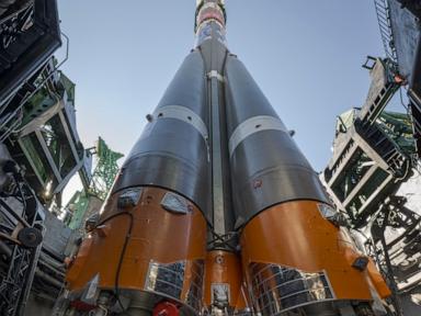
NASA Administrator visits Russia’s Baikonur launchpad for U.S.-Russian ISS crew launch
NASA Administrator Jared Isaacman travels to the Russia-leased Baikonur cosmodrome in Kazakhstan to attend a scheduled l...